Knee Replacement Surgery
what is a knee replacement surgery?
Knee replacement, also called knee arthroplasty or total knee replacement, is a surgical procedure to resurface a knee damaged by arthritis. Metal and plastic parts are used to cap the ends of the bones that form the knee joint, along with the kneecap. This surgery may be considered for someone who has severe arthritis or a severe knee injury.
Various types of arthritis may affect the knee joint. Osteoarthritis, a degenerative joint disease that affects mostly middle-aged and older adults, may cause the breakdown of joint cartilage and adjacent bone in the knees. Rheumatoid arthritis, which causes inflammation of the synovial membrane and results in excessive synovial fluid, can lead to pain and stiffness. Traumatic arthritis, arthritis due to injury, may cause damage to the cartilage of the knee.
The goal of knee replacement surgery is to resurface the parts of the knee joint that have been damaged and to relieve knee pain that cannot be controlled by other treatments.
Anatomy of the knee
Joints are the areas where 2 or more bones meet. Most joints are mobile, allowing the bones to move. Basically, the knee is 2 long leg bones held together by muscles, ligaments, and tendons. Each bone end is covered with a layer of cartilage that absorbs shock and protects the knee.
There are 2 groups of muscles involved in the knee, including the quadriceps muscles (located on the front of the thighs), which straighten the legs, and the hamstring muscles (located on the back of the thighs), which bend the leg at the knee.
Tendons are tough cords of connective tissue that connect muscles to bones. Ligaments are elastic bands of tissue that connect bone to bone. Some ligaments of the knee provide stability and protection of the joints, while other ligaments limit forward and backward movement of the tibia (shin bone).
The knee consists of the following:
-
Tibia. This is the shin bone or larger bone of the lower leg.
-
Femur. This is the thighbone or upper leg bone.
-
Patella. This is the kneecap.
-
Cartilage. A type of tissue that covers the surface of a bone at a joint. Cartilage helps reduce the friction of movement within a joint.
-
Synovial membrane. A tissue that lines the joint and seals it into a joint capsule. The synovial membrane secretes synovial fluid (a clear, sticky fluid) around the joint to lubricate it.
-
Ligament. A type of tough, elastic connective tissue that surrounds the joint to give support and limits the joint's movement.
-
Tendon. A type of tough connective tissue that connects muscles to bones and helps to control movement of the joint.
-
Meniscus. A curved part of cartilage in the knees and other joints that acts as a shock absorber, increases contact area, and deepens the knee joint.
During the procedure
Knee replacement requires a stay in a hospital. Procedures may vary depending on your condition and your doctor's practices.
Knee replacement surgery is most often performed while you are asleep under general anesthesia. Your anesthesiologist will discuss this with you in advance.
Generally, knee replacement surgery follows this process:
-
You will be asked to remove clothing and will be given a gown to wear.
-
An intravenous (IV) line may be started in your arm or hand.
-
You will be positioned on the operating table.
-
A urinary catheter may be inserted.
-
If there is excessive hair at the surgical site, it may be clipped off.
-
The anesthesiologist will continuously monitor your heart rate, blood pressure, breathing, and blood oxygen level during the surgery.
-
The skin over the surgical site will be cleansed with an antiseptic solution.
-
The doctor will make an incision in the knee area.
-
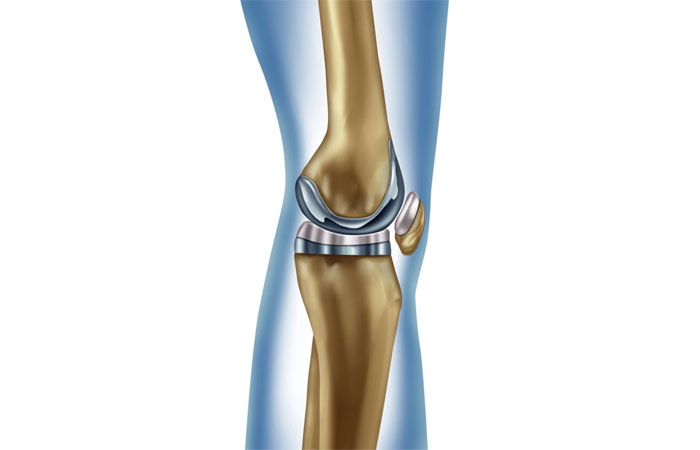
The doctor will remove the damaged surfaces of the knee joint and resurface the knee joint with the prosthesis. The knee prosthesis is made up of metal and plastic. The most common type of artificial knee prosthesis is a cemented prosthesis. Uncemented prostheses are not commonly used anymore. A cemented prosthesis attaches to the bone with surgical cement. An uncemented prosthesis attaches to the bone with a porous surface onto which the bone grows to attach to the prosthesis. Sometimes, a combination of the 2 types is used to replace a knee.
The prosthesis is generally comprised of 3 components: the tibial component (to resurface the top of the tibia, or shin bone); the femoral [thigh bone] component (to resurface the end of the thighbone; and the patellar component (to resurface the bottom of the kneecap that rubs against the thighbone).
-
The incision will be closed with stitches or surgical staples.
-
A drain may be placed in the incision site to remove fluid.
-
A sterile bandage or dressing will be applied.